

year 12 work experience week
Aspiring Year 12 medic Jasmine tells us about her hospital placement in Aintree
As part of our ongoing Careers provision, this week our Year 12 students are engaging in a week of work experience placements in a huge variety of sectors and professions; and next week, it will be the turn of our Year 10 students.
Jasmine 12X, who aims to study Medicine, has successfully secured a placement within an A & E department, a specialism that she is particularly keen to pursue. We are looking forward to hearing more about all that she has learnt this week once Jasmine returns, and here she tells us more about her recent placement in Radiology:
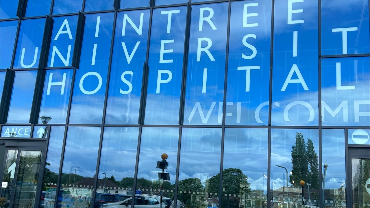
During May's half-term break, I benefitted from a 3-day work experience placement at Aintree Hospital’s radiology department. It allowed me to gain insight into how the hospital works on a day-to-day basis, and also the roles of different people.
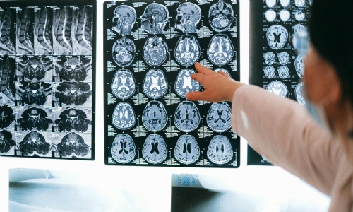
On the morning of day 1, I went to the CT and MRI scan department, where I got the opportunity to observe a biopsy. Biopsy is the extraction of sample cells or tissues for examination to determine the presence or extent of a disease. The patient I saw had a lesion in her brain; although it had not caused any symptoms yet, if it worsened, it might cause damage. So the doctors decided to extract part of the tissue and send it to the laboratory for testing to find out the potential disease.
The whole process takes around 1 hour. This includes the doctor studying the brain scan, giving anaesthetics, confirming the patient's condition, and performing the intervention. The MRI scan shows the image of the brain directly on screen so that the surgeon can observe the position and angle from the scan in order to make sure that the needle is inserted precisely where it needs to be.
Observing the whole procedure helped me to learn and acquire decision-making skills, as the doctor needs to study and assess the patient’s condition to make sure that they are suitable for a biopsy before carrying out the intervention.
For example, another patient had an infection on that day, so the doctor decided to do the biopsy on a later date, as infection clouds the area from which they want to extract, meaning that the MRI scan would be pointless. And it could be potentially harmful if the needle is inserted in the wrong place as a result of a blurred image.
This experience helped me to realise the importance of teamwork and communication, as a biopsy needs to be conducted by a group of workers: a doctor performs the intervention, a radiologist analyses the MRI scan, and a nurse reassures the patient and collects the sample.
In the afternoon, I was present at the radiology outpatient clinic, where people from GP/ fracture clinic are referred to. I saw how patient the staff are, because most of the patients having X rays lacked mobility (eg. a broken leg), so the radiologists carefully adjust their posture in order to get the best image.
The radiologists talked me through the data recording process for an X-ray scan. There is a record of dosage used in each X ray, so that doctors in the future can check the records, and make sure that it doesn’t exceed the limit.
On day 2, I shadowed the doctors in the fluoroscopy department. Fluoroscopy is a type of medical imaging that shows a continuous X-ray image on the monitor. There are two main types performed in the hospital: barium swallows and barium meals. Barium meals look and focus on the stomach, whereas barium swallows look at the oesophagus. It checks for problems in the upper gastrointestinal tract, which includes mouth, back of throat, oesophagus and stomach. By asking the patient to swallow the barium solution and tracking its way through the body, it can help diagnose oesophageal disorders, ulcers, structural problems in the GI tract, and tumours.
In the afternoon, I observed the ultrasound department. One of the patients had a swollen leg and by using an ultrasound scanner, doctors can analyse the blood flow in the legs and blood pressure. This is because the imbalance between osmotic and hydrostatic pressure may be one of the causes of swelling. On the ultrasound scanner, there are different colours covering different areas. The colour scale represents the direction of blood flow, with red meaning blood flowing towards the probe, and blue is blood flowing away from the probe.
This allows doctors to accurately measure the blood flow. There is also a graph of the volume of blood against time. The volume rises every time the heart beats, and the doctor chooses the lowest point and the peak to measure the time between the two points (indicated time of heart contraction). A normal range is lower than 0.07s in order to supply sufficient blood to our body.
Lastly, I observed the administration of injections using ultrasound. Patients had steroid injections into their joint whilst ultrasound is used to monitor the position of the needle. Steroid injections are used to treat problems like joint pain, inflammation, and swelling. This can relieve the redness, pain, and stiffness of the joint.
Before the procedure, doctors ensure that they explain the potential risks of injection, clearly including infection, allergic reaction, and short-term pain. Then they reassure the patient or even distract them by chatting, in order to calm their nerves. This demonstrates the empathy that doctors practise, as they understand the patients’ fear, and try to reassure them.
I learnt a great deal during this 3-day placement, and I am very grateful to have had the chance to observe a variety of departments and procedures.
It is very interesting to see how the hospital operates behind the scenes, and I have acquired and learnt really valuable skills from the experience that will support my plans, aspirations and application for a medical career.
Report and photograph by Jasmine 12X